.jpg?resize=1600x0)
Welcome to the SEMS History
You will find here various fragments of our history and that of the field of microscopy in general. I hope you will find the postings interesting and educational. If you would like to comment on a posting or contribute your own posting you can send the material to me and I will see that it gets posted. Moreover, I welcome any comments or suggestions you might have for the site.
Sincerely,
Jay Jerome
SEMS Historian and Archivist
Brief History of SEMS by Jay Jerome
We began life as the Southeast Electron Microscopy Society (SEEMS). The organizational meeting was held on May 22, 1964 at Emory University in Atlanta, GA. In March of 1964 Dr. Anthony Kattine, then an Assistant Professor of Pathology at Emory, sent out a letter to Universities and Colleges in the Southeast to determine interest in forming a regional organization for electron microscopists. Having received an enthusiastic response, Dr. Kattine announced that an organizational meeting would be held at Emory University on May 22. The meeting was attended by almost fifty scientists from a five state area. It took place from 2:30-5:30 that day in the Woodruff Memorial Building of the School of Medicine. During the meeting, Dr. Kattine was elected Chairman, Mr. John Brown was elected Chairman-elect, and Mr. Ben Spurlock was elected Secretary-Treasurer. Following the meeting, the attendees had a Dutch treat dinner in the Emory University Cafeteria.
The executive committee drafted a constitution and by-laws. Ben Spurlock sent out the minutes of the meeting, the constitution and by-laws, and an announcement that the organization would meet three times a year with Atlanta as the “home base.” Dues for the first year were $3.00 for charter and regular members, $20.00 for corporate members. On October 1, 1964 SEEMS reported 46 regular members, 4 corporate members, and a treasury of $226. The purpose of the new society was stated as follows: “That a society should be formed to facilitate the exchange of ideas and information between people engaged in electron microscopy in this area”.
On October 2, 1964 the first “official” meeting of S.E.E.M.S was held. The Woodruff Building at Emory was again the meeting site. The meeting featured a panel discussion on techniques, a real banquet (rather than the cafeteria) and an after dinner talk by Dr. Lucien Caro of Oak Ridge National Laboratories on “ High Resolution Autoradiography.” The second SEEMS meeting (note lack of periods between letters) took place on January 15- 16, 1965 at the School of Medicine, University of Florida. There were 55 registrants. Among the talks was a demonstration of the new Carl Zeiss EM 9 Electron Microscope.
SEEMS grew steadily. By 1970 there were almost 200 members, and over 500 members by 1980. Shortly after our inception, the number of meetings was reduced to 2 per year and in 1974 we switched to one extended meeting per year.
Despite having a membership that is pretty sure of itself, we never quite solidified our identity. S.E.E.M.S or SEEMS, Southeast or South East. However, in 1986 we reincorporated as the Southeastern Electron Microscopy Society and our leader became the President rather than Chairman.
Although this added some clarity it also coincided with the realization that we had not filed a tax return for 10 years. Some pundits suggest the name change was a mechanism for skirting under the IRS radar. In 1992 we dropped the “Electron” from our name. Since we were back in the IRS’s good graces by then, the name change was assuredly to recognize the growing impact other forms of microscopy were making on scientific inquiry. Although we are now SEMS, we maintain our connection to the past by pronouncing our name with a long “E.”
Another important milestone in our history was the introduction in 1972 of the RUSKA AWARD. The RUSKA is one of our highest honors and it is given to the most outstanding paper presented by a student at our annual meeting. Competition for the award is fierce because the quality of our student presentations is high. The list of past RUSKA winners includes many prominent microscopists. The AWARD honors Ernst and Helmut Ruska who were instrumental in the design of early electron microscopes. Ruska and Knoll’s design for magnetic lenses was incorporated into the first electron microscopes to provide better than 10 nm resolution and Helmut Ruska was one of the first scientists to study biological samples by electron microscopy. Until his death in 1997, it was traditional for Dr. Ivan Roth to present the Ruska award to the winners and review the history of the award. He always took great pride in pointing out that SEEMS honored the Ruskas 14 years before the Nobel Prize committee bestowed the physics prize on Ernst Ruska in 1986 and 17 years before EMSA (now MSA) initiated their own Ruska award (they have since dropped the award). The award was originally to honor Ernst Ruska but when the society queried Professor Ruska for permission he insisted the award honor his brother Helmut as well. Given the contributions of both to the field of microscopy and Helmut’s contributions to biology, it is an extra honor that the award is in both Helmut and Ernst Ruska’s name. The first award was planned for the 1971 meeting at Georgia Tech. However, a blizzard forced the cancellation of that meeting so the first Ruska award was presented to Danny Akin at the 1972 meeting in Gainesville, FL. Plaques similar to the one given Danny Akin were also sent to each of the Ruskas.
Over the years much has changed but much has stayed the same. SEMS remains a vibrant exciting society providing information, education, and comradery to and among microscopists in the Southeast. In preparing this brief synopsis, I used information from many sources in the SEMS Archives. In particular, however, I borrowed heavily from previous work by Danny Akin, Johnny Carson, Ray Hart, and Ivan Roth. I thank them and all others for having taken the time to record parts of our history.
Reflections on the Fourth European Regional Conference on Electron Microscopy.
Raymond Hart
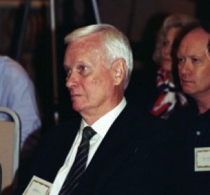
Ray Hart, 1996 Greenville
SC SEMS Meeting
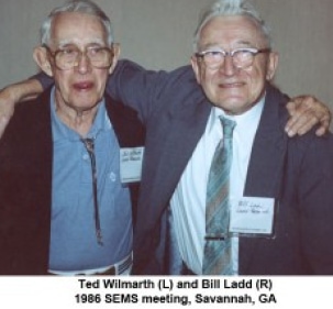
Ted Wilmarth (L) and Bill Ladd (R) 1986 SEMS meeting, Savannah, GA
1965 SEMS MEETING, Atlanta, GA
During the Ruska Award Presentation at the banquet this year (2016), I happened to mention that I am the only SEMS member to have met Ernst Ruska: that meeting occurred during the Fourth European Regional Conference on Electron Microscopy, Rome, Italy, September 1 – 7, 1968.
The Siemens Analytical Instruments Division (SAID) hosted a banquet at the Forum Hotel, which overlooks the Roman Forum and Colosseum. Users of Siemens electron microscopes in North America up to 1968 were invited to that banquet by Cornell W. von Muschwitz, then US director of SAID.
The EMSA (now MSA) members invited to the banquet were Daniel Pease (UC Los Angeles Medical Center), Gareth Thomas (U Berkeley), Robert Hedinreich Bell Labs), Raymond Hart (Argonne National Laboratory) and Albert Crewe (Director, Argonne National Laboratory),
The hosts and more illustrious persons at the banquet were Carla Ruska and Helmut Ruska (Dusseldorf, Germany), Ernst Ruska (Max Planck Gesellschaft, Berlin, Germany), Guston Dupuy (Laboratoire D’Optique Electronique, Toulouse, France), Vernon Cosslett (Cavendish Laboratory, Cambridge, England), and J. B. Le Poole (N.V. Phillips Co., Eindhoven, The Netherlands). The Fourth European conference, to the best of my knowledge was the last major electron microscope meeting that all the above persons were in attendance.
Getting back to Ernst Ruska. I would describe him as a normally soft spoken person with a very clear and precise intellect, and with a gentlemanly manner. He was trained as an Engineer, and of which he was very proud. It is interesting to note that Ernst Ruska was nominated for a Nobel Prize in Physics (because there was no Nobel Prize Award in Engineering) and he turned it down. He did, however, accept the Nobel Prize in Physics in 1986. His co-worker during the late 1920’s and all the 1930’s was B. von Borries, a physicist.
For another reason as to why the 1968 Rome Meeting, considered to be a landmark meeting for electron microscopy, was that it has been described as the divide between “classical electron microscopy” and advanced theory and methodology. Let me clarify that last sentence. Electron microscopy instrumentation up to the late 1950’s and even into the 1960’s was based on theory and practice which was mainly developed prior to the outbreak of WWII.
By the time of the 1968 Rome meeting, the residual gas pressure in a microscope column had been reduced to lower than 10 minus 4 torr and largely purged of carbonaceous debris. Also, the electron beam voltage and lens currents in magnetic lenses had been stabilized to better than 10 minus 4 Amp. Vacuum tubes and other dynamic systems had been largely replaced with solid-state devices. Also, great strides had been made in preparation of specimens for E M. examination: that was especially true in the preparation of biological specimens.
So, when the 1968 Rome meeting convened, microscopists were able to examine their specimens in a clean environment, or a controlled environment, and at a resolution in the vicinity of 2 Angstroms.
Finally, two other electron microscopic methods were actively reviewed at the 1968 Rome meeting. Those methods were scanning electron microscopy/electron dispersive X-ray fluorescence and very high energy (greater than 200 keV) transmission electron microscopy. At the planetary Session on High-voltage microscopy I presented a paper, “Electron Microscopy: The High Voltage Approach”.